Calcific Tendinitis
What is Calcific Tendinitis?
Calcific tendinitis is a build up of a calcium within the rotator cuff tendons. It most commonly occurs in adults between the ages of 30 and 60.
What causes a Calcific Tendinitis?
The cause is unknown but thought to be related to overuse and wear and tear of the tendon. It most commonly occurs in the Supraspinatus tendon which is involved in lifting the arm away from the side of the body.
What are the symptoms and signs?
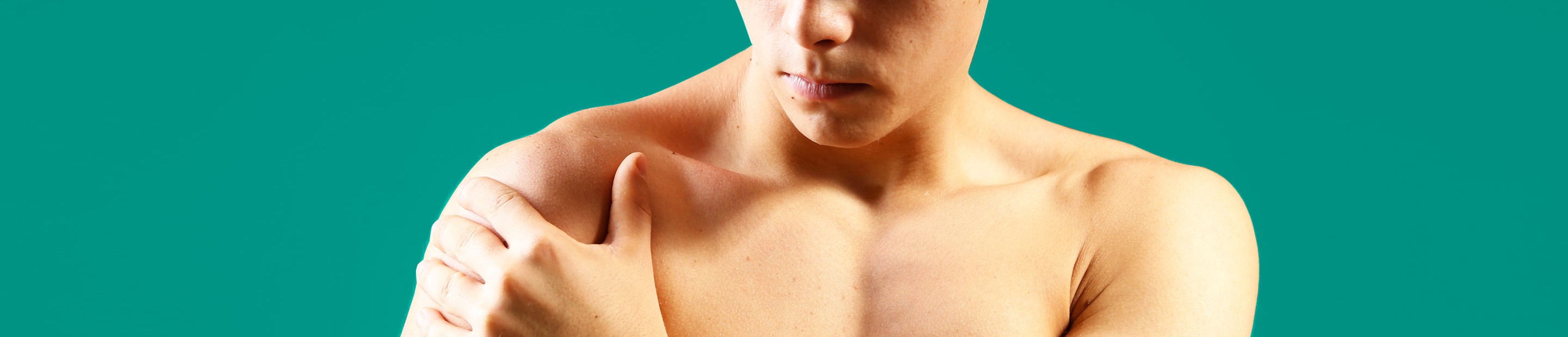
Calcific tendinitis often presents as shoulder pain which can be fairly quick in onset. It may disturb your sleep at night and prevent you putting your arm in certain positions such as reaching out to the side. Because it takes up room in the tendon, it can make the tendon bulkier and cause symptoms identical to those seen in impingement syndrome. Occasionally it can lead to stiffness in the shoulder (Frozen shoulder).
In many patients however, it can occur without causing any symptoms and is found incidentally on an x-ray.
How is it diagnosed?
It is generally detectable using standard x-ray examinations but also can be seen on ultrasound and MRI scans.
What treatments are available?
The mainstay of treatment is through non-surgical means. If left alone, the calcium resorbs over time. This can range from a few weeks to several years.
Pain killers – these are an excellent way to reduce your symptoms during the painful episodes. Paracetamol and anti-inflammatory medications are the best first line and your doctor or pharmacist will be able to advise which are safe for you to take.
Injections – steroid injections into the shoulder joint can be effective in the short term at improving pain. These can be administered by your general practitioner or in the outpatient clinic. This type of injection helps to settle down the inflammation and impingement caused by the calcium deposits.
Barbotage and Aspiration – this technique can be very effective in improving pain and removing the calcium from the tendon. It is performed under ultrasound guidance in combination with local anaesthetic. One or more needles are passed into the tendon to break down the deposit and ultimately remove it through the syringe (aspirate). The area surrounding the tendons can then be injected with steroid to help limit pain after the procedure.
Extra-corporeal Shock Wave Therapy – this treatment can be used on its own or in combination with ultrasound guided aspiration of the calcium. It involves using an external probe on the skin of the shoulder to send minute shock waves into the calcium to break it down.
Surgery – this final option is keyhole surgery to the shoulder. This is performed under general anaesthesia (with you asleep) during which the calcific deposit is localised and then removed from the shoulder. At the same time, the rest of the shoulder undergoes a thorough examination to ensure there are no further abnormalities. This procedure is undertaken as a day case meaning you can normally leave hospital on the same evening. It is normal to experience some discomfort after this procedure until the inflammation resolves. Complications are uncommon but include persistent pain, stiffness/frozen shoulder, recurrence or incomplete excision of the calcium.